Precision Surgery, Miniaturized and Wearable Spectroscopic/Imaging Devices.
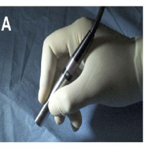
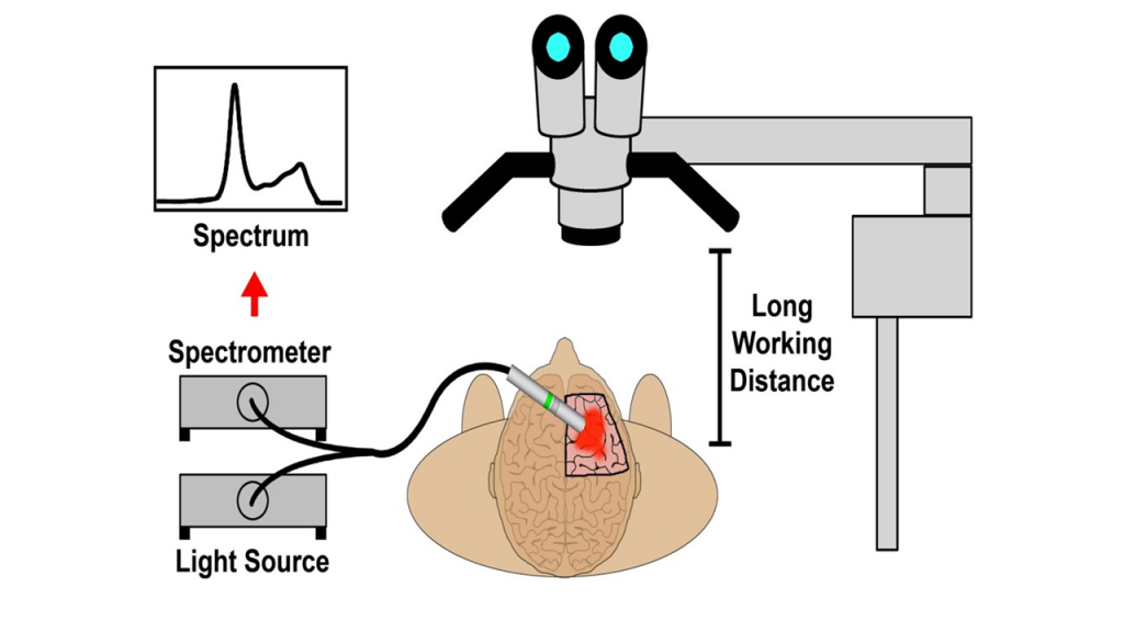
Handheld Spectroscopic Devices – “SpectroPen” Optical Spectroscopy for Image-Guided
Surgery of Malignant Brain Tumors
2) Handheld device development
- Multispectral and multi-modality imaging chips, miniaturized CMOS camera for endoscopic and minimally invasive procedures
3) Imaging probes and targeting tracers
- NIR dyes, quantum dots, SERS nanoparticles, Up-conversion/down-conversion nanoparticles, carbon-dots, and cell-based contrast agents
Single-Molecule and Single Nanoparticle SERS
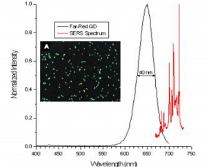
Optical detection and spectroscopy of single molecules and single nanoparticles have been achieved at room temperature with the use of surface Enhanced Raman spectroscopy.
Optimization of SERS Nanoparticle Tags
A new route was developed to increase SERS intensities and nanoparticle stability through hydrophobic domain PEG ligands, achieving highly sensitive detection in biological imaging applications.
4) Augmented reality and artificial intelligence (software)
- Computer vision, tactile and haptic feedback, etc.
5) Clinical and translational studies
- Lymph nodes mapping, intraoperative detection of positive (metastatic) lymph nodes, differentiation of cancer and inflammation
SERS Nanoparticle Tags for In Vivo Tumor Targeting and Intraoperative Tumor Detection
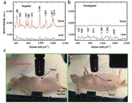
The biocompatible and nontoxic nanoparticles was reported for in vivo tumor targeting and detection based on pegylated Au nanoparticles and SERS.
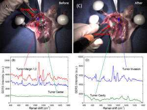
SERS nanoparticles for intraoperative tumor margin detection.
6) Intraoperative mapping and differentiation of nerves. Blood vessels, and other vital tissue studies and organs during robotic surgery
7) Fundamental studies of sensitivity, specificity, light scattering and tissue penetration depth, tumor heterogeneity and cellular and tissue targeting
8) Studies of two near-infrared window (680-950 nm and 1000-1700 nm) for in vivo intraoperative tumor image,afterglow and wavelength-shifting probes for surgical guidance
In-Vivo Imaging and Spectroscopy in Second Near-infrared (NIR-II) Window
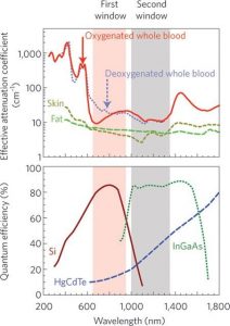
Two clear windows in the near-infrared (NIR) spectrum are of considerable interest for in vivo molecular imaging and spectroscopic detection. The first clear window (650-950 nm), has been shown to be far superior than visible light. The second window (1000–1700 nm), has been reported to further improve the detection sensitivity, spatial resolution, and tissue penetration.
Reference Papers
Nie, Shuming, and Steven R. Emory. “Probing single molecules and single nanoparticles by surface-enhanced Raman scattering.” Science 275.5303 (1997): 1102-1106.
Qian, Ximei, et al. “In vivo tumor targeting and spectroscopic detection with surface-enhanced Raman nanoparticle tags.” Nature biotechnology 26.1 (2008): 83.
Smith, Andrew M., Michael C. Mancini, and Shuming Nie. “Bioimaging: second window for in vivo imaging.” Nature nanotechnology 4.11 (2009): 710.
Lane, Lucas A., Ruiyang Xue, and Shuming Nie. “Emergence of two near-infrared windows for in vivo and intraoperative SERS.” Current opinion in chemical biology 45 (2018): 95-103.