Friday, Feburary 26
15:00 – 18:00
Zoom link in registration email
With advances in biological sensing and imaging techniques and the resulting increase in the availability of biological data, computational tools are becoming increasingly important to life sciences. These tools range from signal processing and machine learning for modelling, to algorithms and computing systems for decision making. The session will bring together researchers and students to discuss the latest advances in the fields of bioimaging and computational biology.
This session will consist of topics including but not limited to the following – (1) computational techniques for biomedical image reconstruction, processing and analysis, (2) computational approaches to structural biology, genomics and neuroscience, and (3) interdisciplinary solutions for bioimaging and life sciences integrating multiple computational approaches and/or hardware and experiments. This session will feature a keynote speech by Prof. Roarke Horstmeyer from Duke University and an invited talk by Ellen D. Zhong from MIT CSAIL.
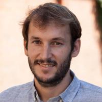
Keynote Speaker – Prof. Roarke Horstmeyer, Duke University

Title: Intelligent Biomedical Imaging Devices
Time: 15:00 – 16:00, Feburary 26
Abstract: Over the past several years, the field of artificial intelligence in general, and machine learning in particular, has transformed how we gain insights from biomedical image data. Now, new machine learning tools are also being used to optimize how we acquire image data. Using deep neural networks to jointly optimize software and hardware is producing a new class of “intelligent” imaging device, which can be designed to probe and inspect in a semi-autonomous manner to yield precise insights, or even create new types of image data. In this talk, I will present some of my lab’s recent work in this new field. I will show examples of a new intelligent microscope that uses patterned illumination to classify infectious disease, or segment cancerous tissue, with higher accuracy than standard microscopes. I will also show how we are using machine learning to create better 3D images in several microscopes. In addition, I will discuss a new approach to map out blood flow within thick (5 mm+) and rapidly decorrelating tissue at video frame rates.
Biography: Roarke Horstmeyer is an assistant professor within Duke’s Biomedical Engineering Department. He develops microscopes, cameras and computer algorithms for a wide range of applications, from forming 3D reconstructions of organisms to detecting neural activity deep within tissue. His areas of interest include optics, signal processing, optimization and neuroscience. Most recently, Dr. Horstmeyer was a guest professor at the University of Erlangen in Germany and an Einstein postdoctoral fellow at Charitè Medical School in Berlin. Prior to his time in Germany, Dr. Horstmeyer earned a PhD from Caltech’s electrical engineering department in 2016, a master of science degree from the MIT Media Lab in 2011, and a bachelors degree in physics and Japanese from Duke University in 2006.
Student Keynote Speaker – Ellen Zhong, MIT CSAIL

Title: Neural reconstruction of protein structure from cryo-EM images
Time: 16:00 – 16:30, Feburary 26
Abstract: Cryo-electron microscopy (cryo-EM) is a powerful experimental technique for determining the structure of proteins and other macromolecular complexes at near-atomic resolution. In single particle cryo-EM, the central problem is to reconstruct the 3D structure of a macromolecule from 104-7 noisy and randomly oriented 2D projection images. However, the imaged protein complexes may exhibit structural variability, which complicates reconstruction and is typically addressed using discrete clustering approaches that fail to capture the full range of protein dynamics. Here, I introduce cryoDRGN, a neural method for cryo-EM reconstruction that extends naturally to modeling continuous generative factors of structural heterogeneity. This method encodes 3D protein structures in Fourier space using coordinate-based deep neural networks, and trains these networks from unlabeled 2D cryo-EM images. We demonstrate that the proposed method can perform fully unsupervised reconstruction of 3D protein complexes from synthetic datasets. On real datasets, cryoDRGN is able to uncover residual heterogeneity in high resolution datasets, reveal new conformations of macromolecular machines and visualize continuous trajectories of their motion. CryoDRGN is open-source software freely available at http://cryodrgn.csail.mit.edu.
Biography: Ellen Zhong is a fourth-year Ph.D. student in the MIT Computer Science and Artificial Intelligence Lab advised by Bonnie Berger and Joey Davis. She is interested in problems at the intersection of computation and biology: her current research focuses on developing neural methods for 3D reconstruction of protein structure from cryo-EM images. Prior to MIT, she was a research associate and scientific programmer at D. E. Shaw Research, where she developed algorithms and infrastructure for predicting protein-small molecule binding affinities from molecular dynamics simulations on Anton supercomputers. She obtained her bachelor’s degree from the University of Virginia, where she worked with Michael Shirts on studying protein folding using Hamiltonian Monte Carlo simulations. She recently co-organized the first workshop on Machine Learning in Structural Biology at NeurIPS, and is a recipient of an NSF Graduate Student Research Fellowship.
Student Speakers
 Grant Greenberg, UofI
Sequence Modelling to Improve Genome Assembly Graph Structure
16:30 – 16:50, Feburary 26
 Xiaohui Zhang, UofI
Automated sleep stage scoring of mesoscale calcium imaging via multiplex visibility graph and deep learning
16:50 – 17:10, Feburary 26
 Al Smith, Carle-Illinois College of Medicine
Neurosurgical Navigation Robot for Low Resource Communities
17:10 – 17:30, Feburary 26
 Vaishnavi Subramanian, UofI
Multimodal Fusion using Sparse CCA for Breast Cancer Survival Prediction
17:30 – 17:50, Feburary 26