
Research
Visualization of cellular machinery by fluorescence microscopy
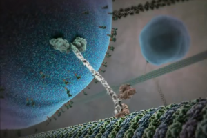
The walking protein: Kinesin protein that walks on cellular roads using its two feet and carries a cargo on it head. Image credits: BioVisions at Harvard.
Proteins, by virtue of their small sizes, are difficult to observe. In one of the techniques, proteins are attached with the specific molecules (called fluorophores) that can ‘light up’ upon shining light of certain wavelength. Dynamics of such ‘lighting up’ molecules, that are attached to the protein of our choice, can be tracked by using certain microscopic components. Kinesin protein is shown in the figure that walks on microtubules (cellular roads) with a constant step size of 8 nm. Walking kinesin was visualized using fluorescence microscopy in the lab of Prof. Paul Selvin and its step size was determined (Yilditz et al., Science). I am using similar fluorescence microscopy techniques for understanding brain function and proteins related to human diseases.
Understanding Molecular Machines
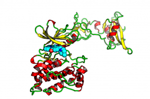
Src Kinase: Important in Cancer Progression
Every part of our body consists of complex networks of machines that are working in co-ordination with each other. These machines are proteins. They are power-horses of life. Whether it be pumping of blood from heart or firing of neurons for the memory; taste of the taste buds or capacity to fight diseases; life processes are driven by proteins. Therefore, it is extremely important to understand how these machines are working.
Proteins are very tiny and very fast. They primarily work by changing their shapes and interacting with other proteins/molecules. Any microscope of the world is not capable to observe the motion of tiny and rapidly changing proteins. I observe the motions of proteins computationally. I use Molecular Dynamics simulations to visualize how proteins are changing their shapes, interacting with other molecules.
Shown is the protein named Src Kinase that regulates cell growth. If its shape does not change the way it should for healthy cell growth, cells start to grow rapidly and without any restraint, a condition, that we call Cancer.
Medicines for Plants
As humans need medicines (drugs) for fighting multiple diseases, agrochemicals are used on plants for productivity and disease/pest resistance. Agrochemicals for plants or drugs for humans, are small organic molecules that stick to certain proteins (in most of the cases) in organisms and change the structure and function of proteins. For example, a drug that binds to Kinase, may help in fighting cancer.
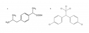
(a) Ibuprofen: Human drug. (b) DDT: Agrochemical (although banned due to its ill effects.)
Interestingly, drugs and agrochemicals look really alike. I am interested in understanding what structural features differentiates a drug from an agrochemical and use machine learning and other statistical approaches.