Dr. Wheeler Among Medical College Inaugural 100 Faculty

DR. WHEELER AMONG MEDICAL COLLEGE INAUGURAL 100 FACULTY

Dr. Wheeler, of the Department of Animal Sciences, is among the 100 prominent researchers, administrators, and medical professionals named to the faculty of the new Carle Illinois College of Medicine, the world’s first engineering-based college of medicine.

The medical college is a partnership between the University of Illinois at Urbana-Champaign and Carle Health System, based in Urbana. The college will welcome its first class of 32 students in 2018.

View the full list of inaugural faculty at the medical college: go.illinois.edu/First100MedFaculty.

Holstein X Gyr F1 Hefiers Arrive at Chessie Creek Farm- Animals Will Be Used to Produce the Next Generation of Tropical Adapted Dairy Cattle

Holstein X Gyr F1 Hefiers

Friday, June 15th, 2018 marked the next step in producing more milk for people in developing tropical countries. Fifty-four Holstein X Gyr F1 heifers arrived at Chessie Creek Farm in South Carolina. The animals are part of the Chessie Creek Farm-University of Illinois Project for the Genetic Improvement of Livestock led by Dr. Matt Wheeler and his colleagues. These animals will provide the genetics for the next generation of embryo donors for this project. Early in the fall some of these animals will calve and the dams will provide the first production data that will ultimately enable the selection of superior genetics to disperse to developing countries. The partnership with Chessie Creek Farm has enabled the fast pace of progress toward the goal of disseminating tropical-adapted dairy genetics worldwide. The University of Illinois wishes to thank the owner and all the staff of Chessie Creek Farm for their support and diligent work on this project.

Matthew B. Wheeler

Gradient Light Interference Microscopy (GLIM) judged one of the ten best microscopy innovations in 2018 by Microscopy Today!

GLIM Imaging for Thick Embryos

Gradient Light Interference Microscopy (GLIM) was judged one of the ten best microscopy innovations in the 2018 Microscopy Today Innovation Award competition. Dr. Marcello Rubessa and Dr. Matthew B. Wheeler and their Beckmann Institute collaborators, Dr. Gabriel Popescu, Dr. Tan Nguyen and Ph.D. student Mikhail Kandel will receive this prestigious award at the Microscopy & Microanalysis 2018 meeting in Baltimore, Maryland, on August 5, 2018.

GLIM extracts extract three-dimensional information from both thin and thick unlabeled specimens. GLIM can potentially become a valuable tool for in vitro fertilization, where contrast agents and fluorophores may impact the viability of the embryo. Since GLIM is implemented as an add-on module to an existing inverted microscope, we anticipate that it will be adopted rapidly by the biological community.

Matthew B. Wheeler

First Tropical Adapted Dairy Calves Born at Chessie Creek Farm in South Carolina!

1/2 Blood Holstein X Gyr Female Calves at Chessie Creek Farm

The first 1/2 blood Holstein X Gyr calves were born in South Carolina in March 2018. These calves will ultimately be the donors that will produce oocytes and embryos for donation to feed developing countries. Chessie Creek Farm is the hub of this work which is a collaboration between Chessie Creek Farm and the University of Illinois through the “Genetic Improvement of Livestock Project”. The project aim is to donate large numbers of Tropical-Adapted cattle embryos to the developing world by 2022. The first donation is being negotiated with Tanzania and will likely occur in the Fall of 2018.

Myogenic potential of mesenchymal stem cells isolated from porcine adipose tissue.

Advances in stem cell biology and materials science have provided a basis for developing tissue engineering methods to repair muscle injury. Among stem cell populations with potential to aid muscle repair, adipose-derived mesenchymal stem cells (ASC) hold great promise. To evaluate the possibility of using porcine ASC for muscle regeneration studies, we co-cultured porcine ASC with murine C2C12 myoblasts. These experiments demonstrated that porcine ASC display significant myogenic potential. Co-culture of ASC expressing green fluorescent protein (GFP) with C2C12 cells resulted in GFP+ myotube formation, indicating fusion of ASC with myoblasts to form myotubes. The presence of porcine lamin A/C positive nuclei in myotubes and RTqPCR analysis of porcine myogenin and desmin expression confirmed that myotube nuclei derived from ASC contribute to muscle gene expression. Co-culturing GFP+ASC with porcine satellite cells demonstrated enhanced myogenic capability of ASC, as the percentage of labeled myotubes increased compared to mouse co-cultures. Enhancing myogenic potential of ASC through soluble factor treatment or expansion of ASC with innate myogenic capacity should allow for their therapeutic use to regenerate muscle tissue lost to disease or injury.

Culture in the presence of differentiating porcine satellite cells can initiate myogenesis in porcine ASC prior to fusion with myotubes. Co-cultures of porcine GFP+ ASC with porcine satellite cells undergoing myogenesis exhibit the presence of individual ASC expressing sarcomeric MHC. a Merged fluorescent images for GFP (green), sarcomeric MHC (red) and dapi (blue) clearly show an individual GFP+ mononuclear cell (arrow) expressing sarcomeric MHC, indicating that this is an ASC that has initiated the myogenic program. Also note the presence of a large GFP+ myotube (*) exhibiting clusters of GFP+ nuclei (arrowhead), indicating that this myotube has been formed with contribution of several ASC. b Enlarged single channel images and merged image of the mononuclear ASC marked by the arrow in (a). The arrow in each panel points to the single nucleus of the cell. Note the presence of GFP label in the nucleus. c Enlarged single channel images and merged image showing a cluster of GFP+ nuclei derived from ASC in the region marked by the arrowhead in (a). The arrow in each panel points to a nucleus derived from a porcine satellite cell lacking GFP

Pictured above: Culture in the presence of differentiating porcine satellite cells can initiate myogenesis in porcine ASC prior to fusion with myotubes. Co-cultures of porcine GFP+ ASC with porcine satellite cells undergoing myogenesis exhibit the presence of individual ASC expressing sarcomeric MHC. a Merged fluorescent images for GFP (green), sarcomeric MHC (red) and dapi (blue) clearly show an individual GFP+ mononuclear cell (arrow) expressing sarcomeric MHC, indicating that this is an ASC that has initiated the myogenic program. Also note the presence of a large GFP+ myotube (*) exhibiting clusters of GFP+ nuclei (arrowhead), indicating that this myotube has been formed with contribution of several ASC. b Enlarged single channel images and merged image of the mononuclear ASC marked by the arrow in (a). The arrow in each panel points to the single nucleus of the cell. Note the presence of GFP label in the nucleus. c Enlarged single channel images and merged image showing a cluster of GFP+ nuclei derived from ASC in the region marked by the arrowhead in (a). The arrow in each panel points to a nucleus derived from a porcine satellite cell lacking GFP

Derek J. Milner, Massimo Bionaz & Elisa Monaco, Jo Ann Cameron and Matthew B. Wheeler


New microscope technique reveals internal structure of live embryos

Marcello Rubessa, Gabriel Popescu and Matthew B. Wheeler teamed up to produce 3-D images of live cattle embryos that could help determine embryo viability before in vitro fertilization in humans.

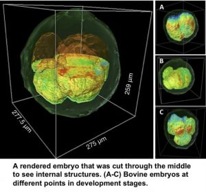
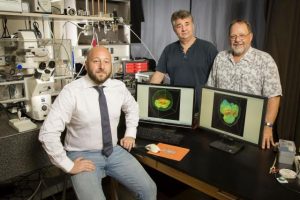
CHAMPAIGN, Ill. — University of Illinois researchers have developed a way to produce 3-D images of live embryos in cattle that could help determine embryo viability before in vitro fertilization in humans. Infertility can be devastating for those who want children. Many seek treatment, and the cost of a single IVF cycle can be $20,000, making it desirable to succeed in as few attempts as possible. Advanced knowledge regarding the health of embryos could help physicians select those that are most likely to lead to successful pregnancies. The new method, published in the journal Nature Communications, brought together electrical and computer engineering professor Gabriel Popescu and animal sciences professor Matthew Wheeler in a collaborative project through the Beckman Institute for Advanced Science and Technology at the U. of I. Called gradient light interference microscopy, the method solves a challenge that other methods have struggled with – imaging thick, multicellular samples. In many forms of traditional biomedical microscopy, light is shined through very thin slices of tissue to produce an image. Other methods use chemical or physical markers that allow the operator to find the specific object they are looking for within a thick sample, but those markers can be toxic to living tissue, Popescu said. “When looking at thick samples with other methods, your image becomes washed out due to the light bouncing off of all surfaces in the sample,” said graduate student Mikhail Kandel, the co-lead author of the study. “It is like looking into a cloud.” GLIM can probe deep into thick samples by controlling the path length over which light travels through the specimen. The technique allows the researchers to produce images from multiple depths that are then composited into a single 3-D image. To demonstrate the new method, Popescu’s group joined forces with Wheeler and his team to examine cow embryos. “One of the holy grails of embryology is finding a way to determine which embryos are most viable,” Wheeler said. “Having a noninvasive way to correlate to embryo viability is key; before GLIM, we were taking more of an educated guess.” Those educated guesses are made by examining factors like the color of fluids inside the embryonic cells and the timing of development, among others, but there is no universal marker for determining embryo health, Wheeler said. “This method lets us see the whole picture, like a three-dimensional model of the entire embryo at one time,” said Tan Nguyen, the other co-lead author of the study. Choosing the healthiest embryo is not the end of the story, though. “The ultimate test will be to prove that we have picked a healthy embryo and that it has gone on to develop a live calf,” said Marcello Rubessa, a postdoctoral researcher and co-author of the study. “Illinois has been performing in vitro studies with cows since the 1950s,” Wheeler said. “Having the resources made available through Gabriel’s research and the other resources at Beckman Institute have worked out to be a perfect-storm scenario.” The team hopes to apply GLIM technology to human fertility research and treatment, as well as a range of different types of tissue research. Popescu plans to continue collaborating with other biomedical researchers and already has had success looking at thick samples of brain tissue in marine life for neuroscience studies. This research was supported by the National Science Foundation, the U. of I. Computational Science and Engineering fellowship and the U. of I. Yuen T. Lo Outstanding Research Award. Editor’s notes: To reach Gabriel Popescu, call 217-333-4840; gpopescu@illinois.edu. To reach Matthew Wheeler, call 217-333-2239; mbwheele@illinois.edu