We select our favorite images every month… well, almost every month… and post the best ones on this website!!!
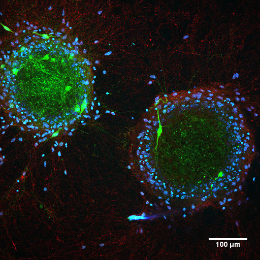 March 2016: Moton neurons (green) from two mouse embryonic stem cell derived embryoid bodies are making synaptic connections as observed using immunostaining of synapsin-1 protein (red). Cell nuclei are stained with DAPI (blue). Credit: Gelson J. Pagán Díaz.
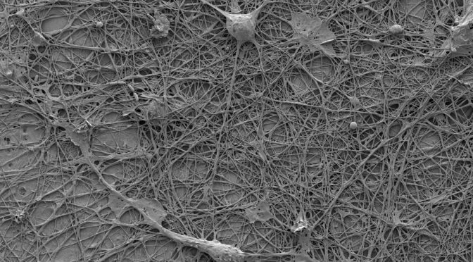
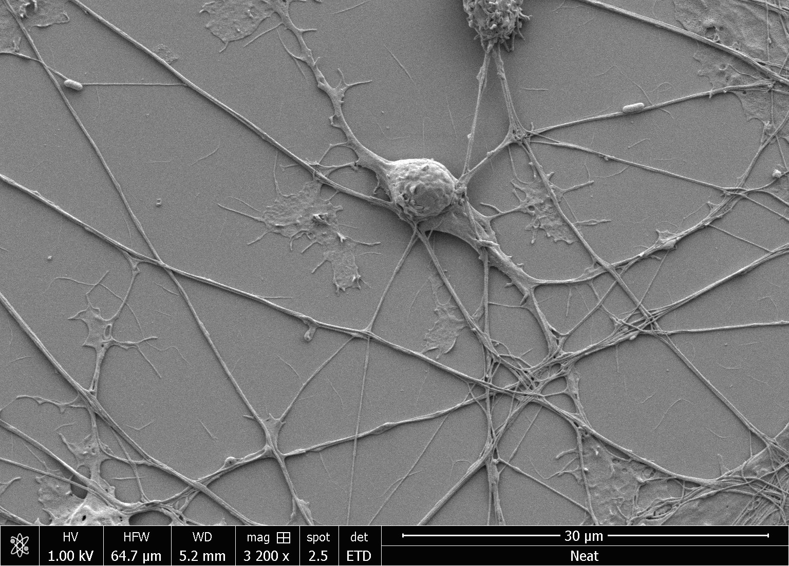 December, 2015: Growth cones of a rat cortical neuron in culture (DIV-09) imaged using scanning electron microscope. Credit: Gelson J. Pagán Díaz.
 September, 2015: Interleukin-1beta (IL-1β) expression in astrocytes (red). Image was recorded from a DIV-16 primary glial culture after immunostaining for IL-1β. Cell nuclei are stained with DAPI (cyan). Credit: Prof. Parijat Sengupta.
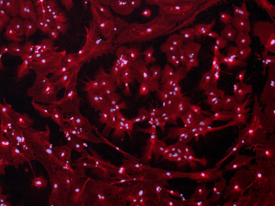
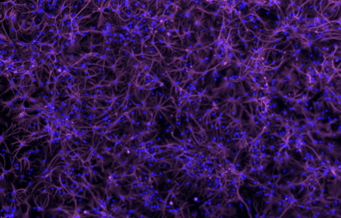 August, 2015: Glial cells immunostained for glial fibrilary acidic protein, GFAP (violet) in a DIV-14 dissociated primary neuron/glia co-culture. Cell nuclei are stained with DAPI (blue). Credit: Prof. Parijat Sengupta.
July, 2015: Light-induced, directional locomotion in muscle ring-powered optogenetic bio-bot with symmetric geometry. This video showed targeted actuation (at 2 Hz) of one muscle ring in a bio-bot. Despite overall symmetry of geometry, the asymmetry in force production causes greater contractile displacement in the stimulated leg (right leg), driving locomotion toward the right. Credit: Ritu Raman and Prof. Parijat Sengupta.