The MRL Central Research Facilities Webinar Series
This free webinar series highlighted techniques available in the MRL Central Research Facilities for the fabrication, processing and characterization of materials. The webinars were free and open to all, even if you are not a current user of our facilities. Webinar topics ranged from very basic, general introductions of techniques all the way to advanced, specialized analyses for featured applications.
Thank you!
Thank you to all the ~2000 people who attended the webinars in this season of the MRL Webinar Series! The MRL central research facilities staff very much enjoyed the opportunity to share our experience with you, focusing on some of our most popular techniques and applications. Recordings of the webinars are available to everyone at go.illinois.edu/MRLYouTubeChannel. Please feel welcome to contact the MRL staff if you have questions or ideas sparked by any of the webinars.
The MRL Central Research Facilities also partnered with the facilities at Penn State University, University of Wisconsin at Madison and University of Minnesota to organize and present another free webinar series covering techniques from all our facilities. MRL staff will be giving talks in that series.
Details here: go.illinois.edu/MRLbig4webinars
Webinars
Top 10 Mistakes in X-ray Analysis (and how to avoid them)
Mauro Sardela
Director, Central Research Facilities, Materials Research Laboratory
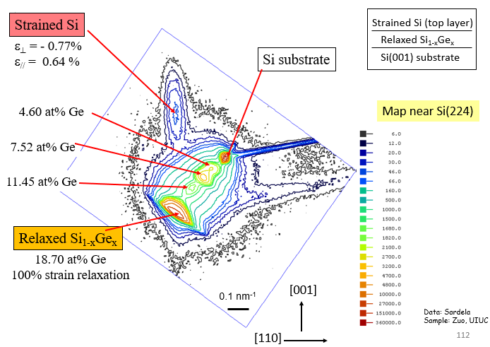
We will present several case studies involving the application of x-ray diffraction analysis in the analysis of materials. Focus will be given to potential artifacts and common mistakes and misconceptions in the technique, ranging from the proper choice of the tool, proper configuration of optics, sample preparation and up to data processing and interpretation. Ten examples will be presented in increasing order of complexity, allowing us to gradually review the basic concepts of the technique and most common analytical methods. Various topics in XRD analysis will be covered: phase identification methods, quantitative analysis of mixtures, preferred orientation, in addition to rocking curve, high resolution scans and reciprocal lattice mapping of thin films as well.
Atom Probe Tomography
Wacek Swiech and Tim Spila, Senior Research Scientists, Materials Research Laboratory
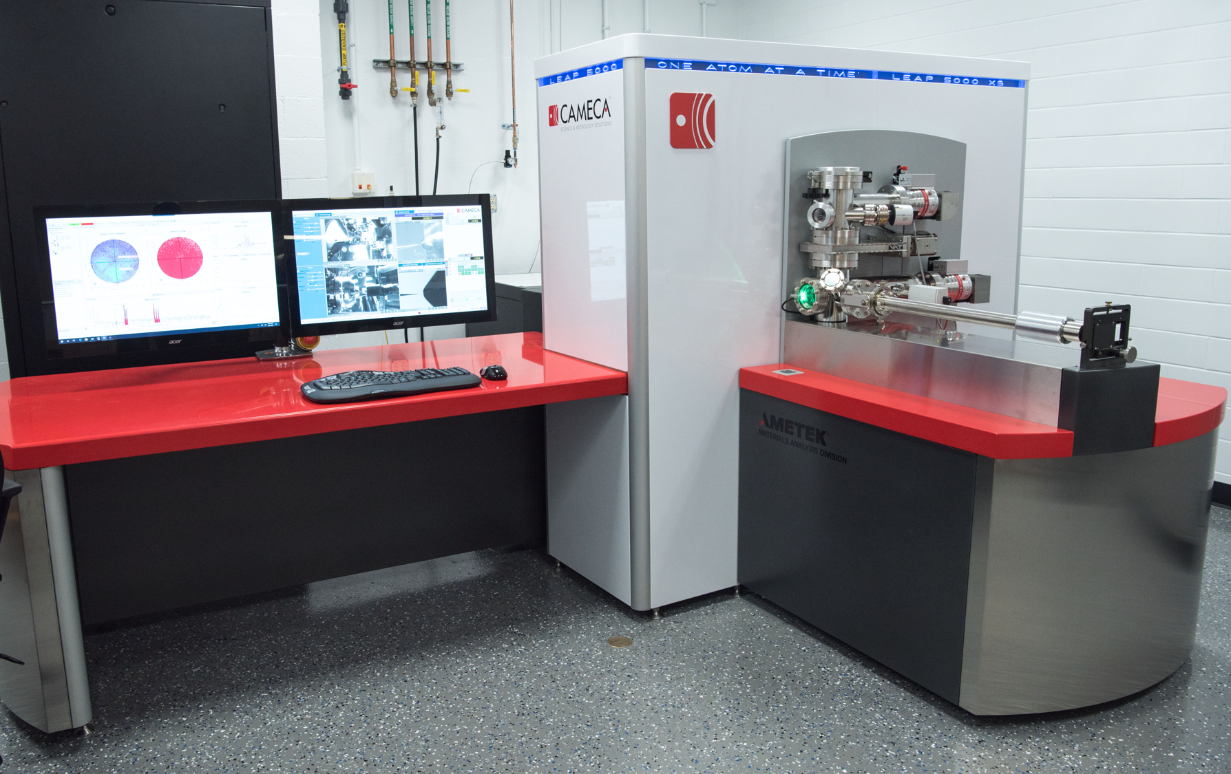 Atom Probe Tomography is an analytical technique where individual atoms are field evaporated from a needle-like specimen in a very high electric field. The ions mass-to-charge ratio is measured on a position sensitive detector allowing for a full, three-dimensional reconstruction of the sample with resolutions less than 0.5 nm for atomic positions. All elements of the periodic table can be analyzed, including isotopic analysis. The APT has been used for grain boundary and interface analysis of metals and thin films to precipitate analysis and imaging of clusters.
This talk will present a brief history of the physics and development of the technique, including work done at the University of Illinois. It will also include an overview of the steps needed to prepare, measure, and analyze a simple sample. Finally, recent examples from Illinois researchers will be presented.
Introduction to Focused Ion Beam
Honghui Zhou
Senior Research Scientist, Materials Research Laboratory
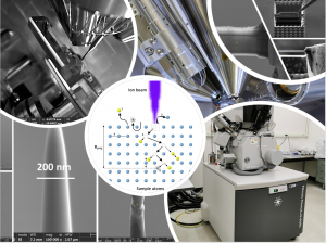 This webinar gives a general introduction to focused ion beam (FIB) technique, which has drawn significant interest among the materials research community due to its unique capability of site-specific milling and depositing at nanometer or micrometer scale. Coupled with scanning electron microscopy (SEM) and equipped with a nano-manipulator and proper analytical detectors, FIB/SEM system can not only image and analyze sample cross-section or hidden features, but also generate a variety of 3 D structures, e.g., site-specific samples for transmission electron microscopy (TEM) and atom probe tomography (APT).
Surface Characterization and Modification of Li-Ion Battery Materials
Rick Haasch
Senior Research Scientist, Materials Research Laboratory
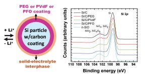 From laptop computers to cellphones, jetliners to racecars, lithium-ion batteries (LIBs) are a part of nearly every aspect of living in today’s world. Over the past decades, the investigation of LIBs has attracted a significant amount of attention. Of importance in the study transition-metal-oxide-based LIBs (TMO-LIBs), is the nature of the interaction of the active oxides with the electrolyte/solvent environment. Many of the issues relating to the performance of TMO-LIBs, such as power fade and capacity loss, are a result of these interactions, with the solid-electrolyte interphase (SEI) playing an important role. The SEI layer provides a protective coating controlling oxide particle wettability, Li-ion transport, electrolyte/solvent oxidation, as well as TM dissolution and migration. This talk will address formation of the SEI layer, its aging, and eventual degradation. Two approaches of cathode surface modification (1) atomic layer deposition (ALD)-alumina-modified lithium nickel cobalt manganese oxide and (2) self-assembled monolayer (SAM)-alkylphosphonate-modified lithium manganese oxide will be presented. Each of these modifications were found to promote Li-ion transport, delay electrolyte/solvent oxidation, as well as reduce manganese dissolution and migration. Characterization methods such as X-ray photoelectron spectroscopy (XPS), secondary ion mass spectrometry (SIMS), and AC impedance spectroscopy will be covered in this talk.
Intro to Small Angle X-ray Scattering (SAXS)
Juan Lopez
Research Scientist, Materials Research Laboratory
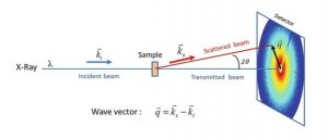 SAXS is a versatile and powerful technique that is often overlooked technique in the materials research community. The purpose of this talk is provide a general introduction to SAXS and its basic analysis. SAXS is an analytical technique used to determine the average size and shape of various particle systems. SAXS is extremely versatile as it can be used to analyze solids, liquids, and gases (captured systems). It is a non-destructive technique and requires little sample preparation. It has applications in a number of different fields such as biological materials, polymers and nanocomposites, just name a few. Variants on SAXS (WAXS, GI-SAXS) will also be covered.
The Versatility of Nanomechanics with AFM
Jessica Spear
Research Scientist, Materials Research Laboratory
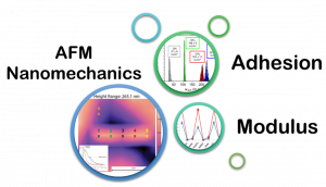 In this webinar, the fundamentals of AFM and force measurements will be reviewed including tip and radius calibration for quantitative measurements. A series of different cases will then be used to show the versatility of nanomechanics with AFM including how to measure adhesion using force curves and functionalized tips, perform adhesion mapping using force volume on 2D materials, measure the stiffness of live bacteria, measure the modulus of stimuli responsive polymers and hydrogels in fluid, and finally highlight unique cases where fast force mapping can provide images of difficult samples not possible with other modes or techniques.
Optical Characterization
Julio Soares
Senior Research Scientist, Materials Research Laboratory
This webinar will give a brief introduction to several modalities of optical characterization of materials. We will offer an overview of each technique covered, through application examples, highlighting their basic principles, instrumentation, modes of operation, possible artifacts, and a few of their limitations and strengths. Among the modalities covered are spectrophotometry (UV-Vis-NIR and FTIR), spectroscopic ellipsometry, Raman spectroscopy, photoluminescence, and nano IR imaging.
Introduction to chemical analysis in (scanning) transmission electron microscopy
CQ Chen
Senior Research Scientist, Materials Research Laboratory
This talk serves as an introduction to chemical analysis in a (scanning) transmission electron microscope (STEM/TEM) with high spatial resolution. I will briefly review the imaging techniques, i.e., Z-contrast imaging and most recently emerged iDPC imaging, for chemical species. I will then transition to the spectra-based techniques, specifically the X-ray energy dispersive spectroscopy (XEDS) and electron energy loss spectroscopy (EELS), which are highly complementary analytical techniques utilizing signals generated by characteristic inelastic scattering of electrons. The basic principles, instrumentation, collection efficiency, capabilities, modes of operation, artifacts, and sample requirements will be discussed.
Electron Microscopy for Biological Materials
Kristen Flatt
Postdoctoral Associate, Materials Research Laboratory
Electron microscopy is a powerful tool that allows us to overcome the limitations of light to investigate the fine structures that make up the world around us. Electron microscopy can uncover many wonders of biology, including the ultrastructure of cells and tissues, morphology and components of infectious viruses, individual proteins and protein complexes, and much more! In this talk, I will give an overview of sample preparation for biological materials and various electron microscopy techniques, including cryo-electron microscopy, with an emphasis on techniques and instruments available here at the MRL. My goal is to highlight the importance of traditional electron microscopy techniques for biological materials research, while also introducing important and exciting new techniques that are spearheading breakthroughs in medical and biological research.
Energy Dispersive X-Ray Fluorescence (ED-XRF) Spectroscopy: Theory and Applications
Mohammad Ali
Postdoctoral Associate, Materials Research Laboratory
Energy dispersive x-ray fluorescence (ED-XRF) spectroscopy is a non-destructive analytical technique, which is used to obtain elemental information of an unknown sample. This technique virtually does not require any sample preparation. Samples can be in powder, thin film, bulk or liquid form. As a result, it is widely employed analytical technique in many industries and applications including cement production, petrochemical, food products, polymers, pharmaceutical, and healthcare products. This webinar will cover the basics of energy dispersive x-ray fluorescence spectroscopy, sample preparation and data analysis.
Thin Film Deposition Techniques-A Comparative Overview
Lon Westfall and Fubo Rao
Research Engineers, Materials Research Laboratory
In this webinar, a variety of thin film deposition techniques and instruments, such as sputtering, evaporation, atomic layer deposition, Parylene deposition, for fabrication of micro- and nano- devices will be described with emphasis on those available at the Materials Research Laboratory. Basic fundamentals of each technique and their pros and cons in depositing films with varied properties will be discussed. We hope to provide qualitative guides for selection of each technique for different applications.
Introduction to Transmission Electron Microscopy
Wacek Swiech, Ph.D.
Senior Research Scientist, Materials Research Laboratory
Transmission electron microscopy (TEM) is the oldest imaging technique using charged particles optics. It has lateral resolution well surpassing the traditional light microscopy. Foundations and basic concepts of conventional TEM will be introduced and discussed. Examples of TEM application not only limited to imaging modes will be shown. Namely the diffraction modes of operation will illustrate the contribution. Additionally, some aspects of scanning TEM (STEM) will be presented. Details of modern TEM/STEM instrumentation equipped with lens aberration correctors resulting in the resolution down to approx. 50 pm will be discussed.
Micro and Nano-lithography
Jeff Grau and Tao Shang
Research Engineers, Materials Research Laboratory
Micro and Nano Lithography is one of the essential steps in Micro/Nano-Fabrication which enables structures, devices and systems in both micrometer and nanometer scales.
Traditional lithography predates the invention of semiconductor devices but it did not fundamentally change the world we see today until photolithography was widely used in the semiconductor industry.
In this webinar, the complete procedure of photolithography is covered step by step. Novel lithography such as maskless laser lithography, Nano 3D Printing and electron beam lithography are also introduced to the audience.
Comparison of different instruments housed in MRL and advice on how to choose one technique over another are offered to audience too.
Practical Microanalysis Based on Scanning Electron Microscopy
Jade Wang, Ph.D.
Research Scientist, Materials Research Laboratory
This talk is focusing on the analytical techniques based on SEM covering from basics to operation. Practical data are used for demonstrating the principles and laws, signal detection and processing, artifacts and other practical aspects in operation and analysis. The presentation will be comprehensive and detailed on the X-Ray microanalysis in SEM and Cathodoluminescence (CL). Diffraction based techniques such as Electron Channeling Contrast microanalysis of defects and practice for getting optimal Electron Backscatter Diffraction (EBSD) data are also briefly discussed.
3D optical profilometry
Julio Soares, Ph.D., and Kathy Walsh, Ph.D.
Sr. Research Scientists, Materials Research Laboratory
With advances in confocal optical microscopy and processing software, 3D optical profilometry became a versatile and easy-to-use technique for measuring surface topography. The technique is complementary to AFM and stylus profilometry for measurements of surface roughness and sample profiles. In addition, 3D optical profilers based on confocal microscopy can produce fully focused optical micrographs of samples with features taller than the depth of focus of the microscope.
Introduction to Secondary Ion Mass Spectrometry
Tim Spila, Ph.D.
Sr. Research Scientist, Materials Research Laboratory
Secondary Ion Mass Spectrometry is an analytical technique for solid materials where a primary ion impinges on a surface causing secondary ions and molecules to be sputtered away. The secondary ions and molecules are then analyzed for their mass-to-charge ratio. Output from the technique comes in the form of mass spectra, surface mass imaging, atomic depth profiling, and 3D atomic depth profile imaging. Information on elemental concentrations can be determined from atomic percent to parts-per-million or part-per-billion for certain elements. All elements of the periodic table can be analyzed, including isotopic analysis. Examples of SIMS analysis of from semiconductor samples, biological materials, wear tested metals, and Li ion batteries will be shown.
Introduction to X-ray photoelectron spectroscopy (XPS)
Rick Haasch, Ph.D.
Sr. Research Scientist, Materials Research Laboratory
X-ray photoelectron spectroscopy (XPS), also known as electron spectroscopy for chemical analysis (ESCA), is a widely used materials characterization technique belonging to the general class of methods referred to as surface analysis. This non-destructive technique provides, to varying degrees, semi-quantitative elemental, chemical-state and electronic structure information from the top 5-10 nm of a material and are sensitive to elements Li and above. XPS has found applications over a vast range of material classes; such as metallic, ceramic, polymeric, and composite; and technologies such as microelectronics, energy conversion and storae, and nanotechnology. Modern spectrometers are now not only capable of achieving high-energy resolution spectroscopy but are also capable of 2-dimensional imaging.
Scanning electron microscopy
Honghui Zhou, Ph.D.
Sr. Research Scientist, Materials Research Laboratory
This webinar focuses on advanced SEM imaging with instrument functions that are available at MRL but may not be well known to our SEM users. Topics covered will include ultrahigh resolution imaging, low voltage imaging and backscattered electron imaging.
Soft materials characterization: an instrument overview
Roddel Remy, Ph.D.
Research Scientist, Materials Research Laboratory
While a plethora of techniques can be used to characterize soft materials, some methods are more commonly associated with the field. This webinar will cover the basics of chromatography, thermal and mechanical analyses, as well as a sprinkling of elastic light scattering, as they relate to soft materials research.
Basics of Atomic Force Microscopy
Kathy Walsh, Ph.D.
Sr. Research Scientist, Materials Research Laboratory
Atomic force microscopy is a versatile technique for looking at surfaces, an excellent complementary technique to SEM and 3D optical profilometry. But it’s more than just pretty pictures! AFM can give insight into mechanical, electromagnetic, or chemical properties of a surface, as well as giving highly precise topographic measurements. This talk will introduce the basics of AFM and will highlight a small number of applications.