
Nanotheranostics
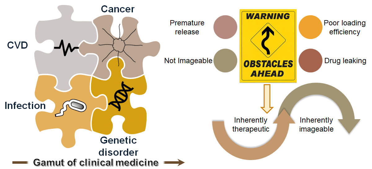
Nanoparticles indeed can be a panacea for a variety of diseases such as cardiovascular disease, cancer, infection and genetic disorder and one can claim the gamut of nanomedicine can be as wide as the clinical medicine. Even though many of the nanoparticles have shown promise in drug delivery, there are obstacles in their translation to bedside for instance poor loading efficiency, drug leaking and premature release. On the top of that, many of the drugs lack inherent imageable signal. My research proposes an alternative approach of utilizing nanoparticles which are inherently therapeutic and imageable.
The main goal of my research is to integrate materials science, advanced materials characterization and biomedical engineering to (1) develop smart nanomaterials for biomedical imaging for the early diagnosis of diseases; (2) develop smart biomaterials for drug delivery applications; (3) assess the safety of the nanomaterials for the ultimate translation from bench to bedside.
Investigation of these smart nanomaterials will not only boost our capability of diagnosis of the diseases at early stage, but also will advance our understanding of the mechanism involved in the progression of diseases while will offer a solution to some of the deadliest diseases in the world.
In other words, my research can be rephrased as ‘find, fight and follow’ where the nanoparticles can detect the disease at early stage, can therapeutically target and treat disease and finally can be used to evaluate the outcome of the therapy.
I. Development of inorganic nanoparticles for computed tomography:
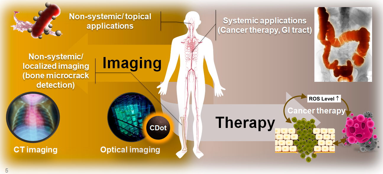
The mainstay diagnostic tool for the assessment of the anatomical feature in clinic is radiographic examination. Contrast agents are used in clinical settings to enhance the tissue visualization on radiographs or computed tomography (CT). Recent developments in the application of nanoparticles (NPs) comprising high atomic number elements have facilitated accomplishing this goal. Throughout my Ph.D. at the University of Illinois Urbana Champaign, I developed nanoparticles based on the high Z elements which carries benefit over the conventional materials used previously. This project is conducted among bioengineering department, veterinary medicine department, Beckman Institute, etc at UIUC and was supported financially by American Heart Association through a $50 K predoctoral fellowship for my Ph.D. studies. In this project, I have been the main coordinating researcher of multidisciplinary research group including vetenrians, 3 undergraduate mentees. These nanoparticles are tuned to detect and cure early infection, bone abnormalities, and GI tract imaging.
II. Development of nanoparticles for optical based imaging:
Due to the high sensitivity of the optical imaging methods and the application of non- ionizing radiation to image and treat diseases, they are envisioned to find a special place in the clinic. However, the interferences from the human organs’ themselves are posing difficulty in differentiating the diseased site. The development of the nanoparticles which can target certain biomarkers while being imageable can introduce a noninvasive strategy for the early diagnosis. Currently, carbon dots are heavily investigated for replacement with the conventional quantum dots and dyes due to their great photoluminescence properties, non-quenching fluorescence signal and most importantly biocompatibility. During my Ph.D. I have worked extensively in two areas of photophysical understanding of carbon dots and their biomedical applications for imaging and drug delivery.
- Fluorescence tuning of carbon dots: Carbon dots were discovered only about a decade ago and therefore; their properties are still quite unknown. Specially, with a surge of interest in the optical based imaging techniques for biomedical applications, conventional quantum dots were rendered ineffective because of their high cytotoxicity. Carbon dots are promising candidates to replace quantum dots due to their unique photoluminescence properties, but their fluorescence tuning is currently highly elusive. During my Ph.D., I have endeavored to elucidate the mechanisms involved in the fluorescence and fine tuning it by changing the composition as the sole driver of the photoluminescence. I have been able synthesize multicolor carbon dots by the controlled chemistry and make their quantum yield close to those of quantum dots.
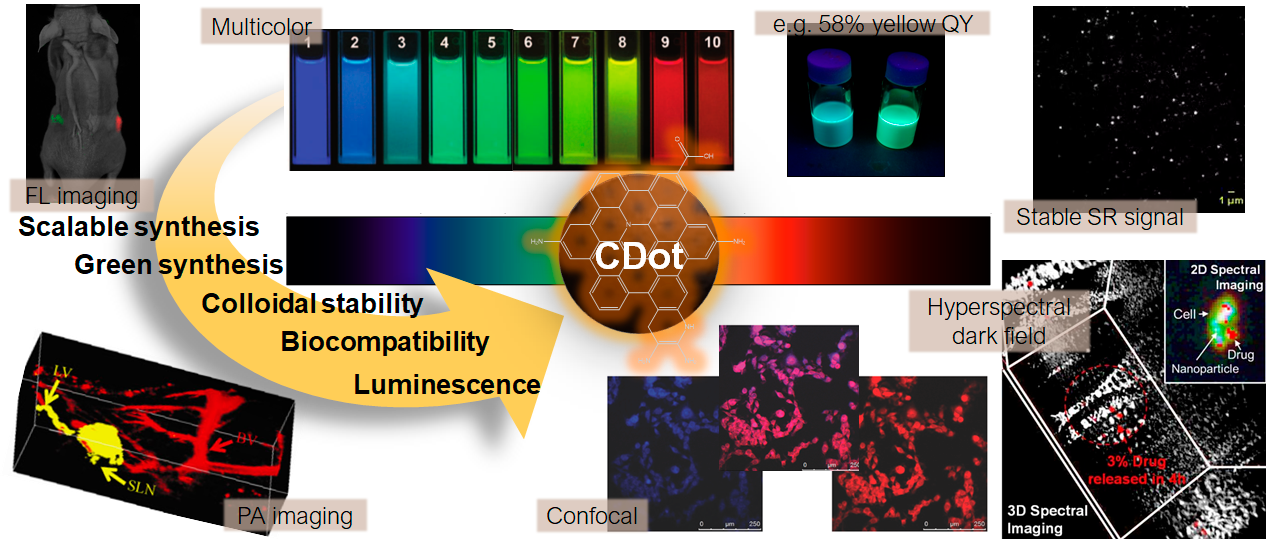
- Biomedical applications of carbon dots: As mentioned above, carbon dots are envisioned to replace conventional quantum dots and hence they can find application where quantum dots were formerly used. In my Ph.D. I applied these nanoparticles as trackable agents in cells by novel hyperspectral darkfield imaging and utilized them for quantification of the drug released in the cells. Moreover, I used these nanoparticles to indicate bone microcrack histopathological features. I also used these nanoparticles as vibrational spectroscopy responsive carriers for the controlled delivery of an anticancer drug in vivo.