Why is printing a 3D Heart from a patient’s MRI or CT scans useful?
There have been several cases in which a surgery was designed based on a 3D printed model. In some cases, the surgery was thought to be inoperable, until the 3D model helped surgeons to plan an alternative access point. http://www.cnn.com/2015/10/06/health/3d-printed-heart-simulated-organs/
3D Printing can allow surgeons to design patient-specific surgeries which could lead to smaller incision sites, better access, reduced surgery time and if a graft or device is being inserted, it could lead to a better fitting model/device. Patient-specific surgeries are very useful in the congenital heart disease community. Patients with congenital heart disease have unique anatomies and complex physiological systems. Surgical planning is often critical to a successful surgery in this patient population. Surgeons can even practice surgeries on the 3D printed models. 
Models can also be used for patient, provider, and medical student education. It is much easier to understand the physiology of an organ when you can hold it in your hand, versus scanning through images on a computer. http://www.jumpsimulation.org/innovation/3d-heart-library.html
A hub for 3D printing from medical images is the NIH 3D print exchange: http://3dprint.nih.gov/
The NIH has created a library of different medical models. These models will be able to make large morphological comparisons possible.
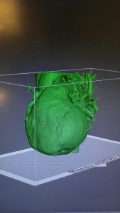
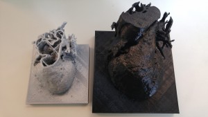
For my semester project, I was able to successfully print a heart from MRI images using Osirix and Meshmixer. Here is a picture of the final project.
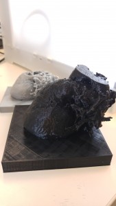
In the future, I hope to write a full tutorial of how to print the internal structures of the heart and how to optimize the printing supports in an extruder print setting.