After literally hundreds of years of investigation the structure of mtitoic chromosomes and the folding motifs giving rise to mitotic chromosome condensation remain poorly understood. Various models have been proposed, each focusing on specific aspects of chromosome structure or molecular features of chromosomal proteins but also extrapolating beyond what is actually known. Serious technical difficulties hinder progress. The size scale of mitotic chromosomes is inconvenient with regard to current structural methods, being too small for conventional light microscopy yet too large for conventional electron microscopy. The packing density is extremely high, meaning that to trace the actual path of the underlying DNA nucleofilament would require ~ nm resolution over a several micron extent. Finally, the chromatin structure is extraordinarily sensitive to small variations in isolation condtions.
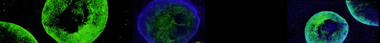
Whereas most approaches have focused on maximally condensed metaphase chromosomes, we have instead pursued the hypothesis that mitotic chromosome condensation occurs through a folding/unfolding continuum beginning in G2, reaching a maximum condensation, and then decondensing through telophase and G1. We have shown that early stages of mitotic chromosome condensation are consistent with a folding of chromonema fibers to form the initial early prophase chromatid which has a diameter roughly half that observed for the metaphase chromatid. We have also shown that later stages of chromosome decondensation in telophase and early G1 are consistent with an unfolding or straightening of chromonema fibers. Finally we have shown that mitotic “scaffold” proteins topoisomerase 2 and SMC2, a component of the condensin complexes, do not assume an axial distribution until later stages of mitotic condensation. At earlier stages, even when a uniformly condensed, linear prophase chromatid is already clearly visible, these proteins are not located at the chromosome axis but rather appear by light microscopy do be localized towards the periphery of the chromatid. Based on these results we have proposed a folded chromonema, axial glue model of metaphase chromosome structure.
Despite our model, we are convinced that major aspects of mitotic chromosome condensation are still simply not understood with different proposed models illuminating different and incompletely understood pieces of the puzzle. Currently we are using engineered chromosome regions as to provide more detailed information on how DNA is folded within mitotic chromosomes, probing aspects such as the reproducibility of chromatin folding within metaphase chromosomes. We this information as providing valuable constraints and tests for any model for chromosome condensation. We are also initiating a closer structural focus on the dynamics and organization of chromosomal proteins such as condensins during the process of mitotic chromosome condensation.
Selected Publications:
Strukov YG, Belmont AS. Mitotic chromosome structure: reproducibility of folding and symmetry between sister chromatids. Biophys J. 2009 Feb 18; 96 (4) :1617-28. PubMed PMID:19217877; PubMed Central PMCID: PMC2717231.
Belmont AS. Mitotic chromosome structure and condensation. Curr Opin Cell Biol. 2006 Dec; 18 (6) :632-8. PubMed PMID:17046228.
Kireeva N, Lakonishok M, Kireev I, Hirano T, Belmont AS. Visualization of early chromosome condensation: a hierarchical folding, axial glue model of chromosome structure. J Cell Biol. 2004 Sep 13; 166 (6) :775-85. PubMed PMID:15353545; PubMed Central PMCID: PMC2172117.
Strukov YG, Wang Y, Belmont AS. Engineered chromosome regions with altered sequence composition demonstrate hierarchical large-scale folding within metaphase chromosomes. J Cell Biol. 2003 Jul 7; 162 (1) :23-35. PubMed PMID:12835314; PubMed Central PMCID: PMC2172725.
Belmont AS. Mitotic chromosome scaffold structure: new approaches to an old controversy. Proc Natl Acad Sci U S A. 2002 Dec 10; 99 (25) :15855-7. PubMed PMID:12461163; PubMed Central PMCID: PMC138527.
Dietzel S, Belmont AS. Reproducible but dynamic positioning of DNA in chromosomes during mitosis. Nat Cell Biol. 2001 Aug; 3 (8) :767-70. PubMed PMID:11483964.
Belmont AS, Bruce K. Visualization of G1 chromosomes: a folded, twisted, supercoiled chromonema model of interphase chromatid structure. J Cell Biol. 1994 Oct; 127 (2) :287-302. PubMed PMID:7929576; PubMed Central PMCID: PMC2120203.