We are interested in new radiation detection and instrumentation for imaging and sensing applications. This includes the development of novel detector technology and imaging techniques, data processing, experimental validation, modelling, computational problem solving, and quantitative characterization of biological processes.
Here are our ongoing projects:
- Design study of a two-panel head and neck dedicated positron emission tomography system
- Penalized MLE image reconstruction for a dual-panel dedicated head and neck PET system
- Autonomous source detection system for anomaly detection and source localization
- Mobile sensor networks for background radiation modeling
Here are our past projects:
- Molecular Imaging
- Modeling and Simulation for Nuclear Imaging
- X-ray Imaging
- Optical and UV detectors
Ongoing projects
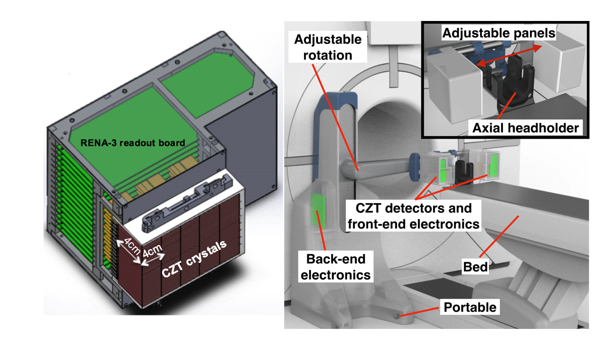
Design study of a two-panel head and neck dedicated positron emission tomography system
We studied the performance of a two-panel positron emission tomography (PET) system dedicated to head and neck cancer (HNC) using Monte Carlo simulation. The goal of HNC treatment is not only to improve patient survival rate but also to preserve organ function and a HNC dedicated PET system with superior performance will help oncologists make better treatment plans. Compared with a commercial wholebody PET system (GE Discovery MI), the proposed dedicated system shows better performance in terms of noise equivalent count rate, photon coincidence sensitivity, spatial resolution and lesion visualization. The NEC rate peak is about 8.9 kcps, and it increases to 10.4 kcps with a lead shielding. The photon sensitivity is 0.89% for a point source placed in the FOV center, and the average sensitivity is 0.64%. With a 2 mm FWHM DOI resolution, the system achieves 1 mm orthogonal-plane and 1.5 mm in-plane spatial resolution. For recovering multiple scattering events, the sensitivity has an approximate 89% sensitivity improvement when recovery correct rate is about 80%, and a 57% improvement when correct rate is about 90%. For lesion visualization, the dedicated system can detect a 2 mm diameter hot rod within 70 s, and SNR and CNR of the reconstructed image get improved after incorporating recovered multiple scattering events. All the performances are better than a state-of-the-art whole body PET system.
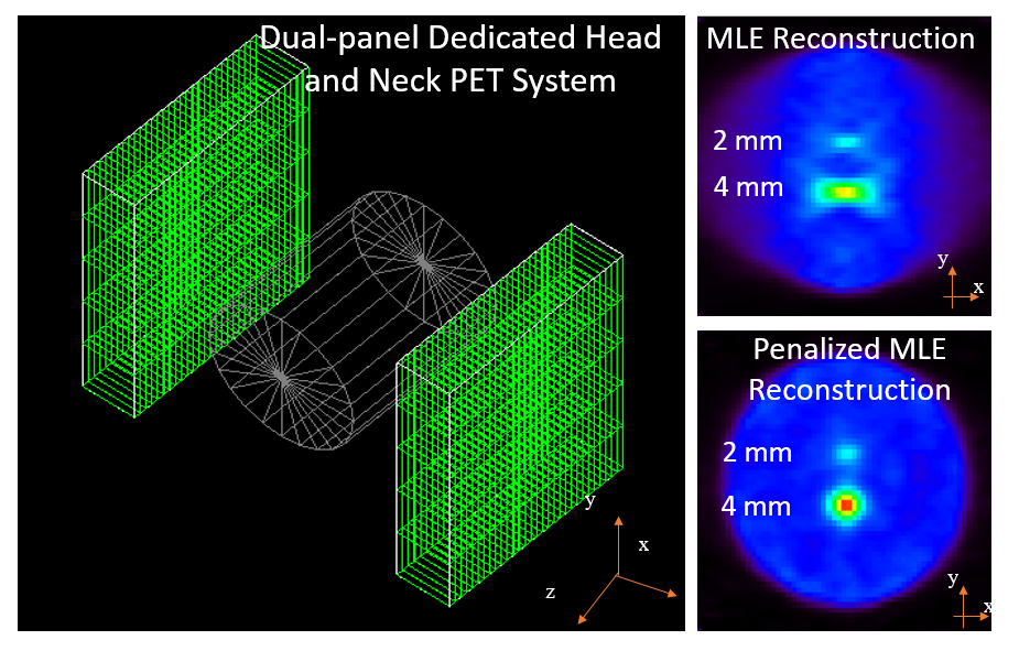
Penalized MLE image reconstruction for a dual-panel dedicated head and neck PET system
The dual-panel system features patient’s comfort, with the detector panels on both sides of the patient’s head and neck. The problem is that the limited angular coverage of the imaging plane leads to artifacts in the reconstructed images, such as the elongation of lesions. We are working on reducing the artifacts by using penalized maximum-likelihood reconstruction.
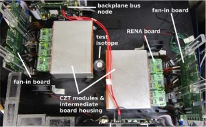
Autonomous source detection system for anomaly detection and source localization
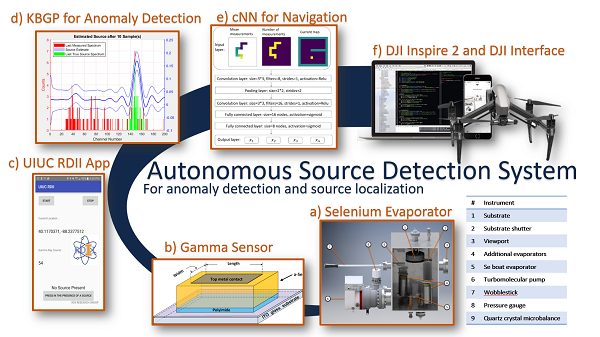
Proliferation of special nuclear material (SNM) is an ever present concern, and technologies for detecting and localizing rogue sources are needed in increasing quality and accessibility to combat potential threats. Many urban settings are left without any security options due to the inhibitive costs and complexity of many detection and localization systems. Our work strives to provide a cost-effective and simple tool for anomaly detection and source localization through development of an autonomous, mobile, single-detector system. The following figure summarizes the many facets of this project below. This is achieved through development of two learning algorithms optimized for sparse data collected from a single, mobile detector, and the construction of an in-house detector that maintains high sensitivity while mitigating cost and weight.
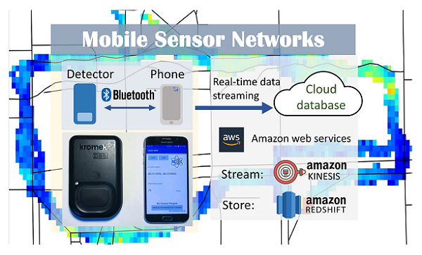
Mobile sensor networks for background radiation modeling
Modeling of background radiation for the urban environment plays an important role in homeland security. However, background radiation is difficult to assess due to its spatial-temporal fluctuations caused by variations in soil composition, building materials, and weather patterns etc. To address the challenge of background radiation modeling, we have developed a mobile sensor network to continuously monitor the background radiation; we have also proposed a maximum likelihood estimation algorithm to decouple and estimate the background’s spatial distribution and temporal fluctuation. Experiment results demonstrate that this background radiation monitoring system accurately recognizes high background regions in the experiment area and successfully captures temporal fluctuation trends of background radiation during rains. Our system provides an efficient solution to model the temporal fluctuation and spatial distribution of background radiation.
Past projects
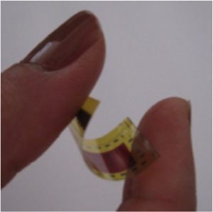
Molecular Imaging
Molecular imaging enables the visualization, characterization, and quantification of biological processes. In nuclear medicine, an instrumental molecular imaging modality is positron emission tomography (PET). We are interested in investigating different detection materials, readout strategies, and algorithms to achieve high resolution, high sensitivity PET.
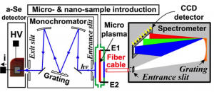
Modeling and Simulation for Nuclear Imaging
We use modeling for validating experimental measurements as well as exploring and evaluating new algorithms for data processing.
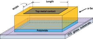
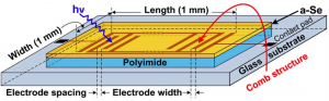
X-ray Imaging
We aim to develop new detector technologies that will provide x-ray images with superior image quality for improved detection of early cancers while also reducing patient risks by increasing detector sensitivity to low levels of radiation.