Nano- and micro-scale platforms for analyzing cell-ECM interactions: collective cell migration, chemoprotective adhesion, and tumor cell invasion.
Cellular interactions with the extracellular matrix (ECM) are important to a number of biological processes, both in healthy and diseased cells. In this talk, I will discuss recent cell-ECM developments in Prof. Dr. Joachim Spatz’s New Materials and Biosystems group at the Max Planck Institute for Intelligent Systems. Concerted collective migration of epithelial cells, observed during wound healing, morphogenesis, and cancer, requires interaction with both the matrix and surrounding cells. A molecular mechanotransduction mechanism governing this process was identified in which the tumor suppressor protein merlin translocates from the cell periphery to the cytoplasm in a Rac1 dependent fashion during migration initiation. Using a micron-scale PDMS trench and a substrate stretching device, this translocation was observed following the application of force in non-migrating cells, suggesting that merlin is a force transducer.
Cancer cells have been found to obtain chemoprotection from certain integrin-ECM interactions. A platform designed to identify and perturb protective matrix properties in a high-throughput manner was built, allowing for the conversion of chemoresistant cells into chemosensitive ones. Block copolymer micelle nanolithography (BCML) was utilized to create scalable arrays of gold nanoparticles to understand the influence of mechanical matrix properties in parallel with ligand presentation on 2D substrates and inside 3D microchannels.
Metastasizing cancer cells must escape their tumor environment and invade surrounding stromal tissue, combining ECM degradation with physical force generation and cytoskeletal rearrangement. PDMS microchannels fabricated with photolithography and replica molding were used to examine the interaction of cancer cells with confined three dimensional environments. A focus was placed on understanding the variations in invasion behavior between cancer cell lines of different tissue origin, then attempting to explain these differences by cell mechanical properties and gene expression. Distinct differences in invasion characteristics were observed between wide 10 μm channels and narrow 3 μm channels, with the latter exhibiting smooth velocity profiles and increased channel permeation. Live cell actin staining, ECM protein alteration, and chemical inhibition of both Rac1 and RhoA mechanical pathways were all used to better understand these differences.
Bio
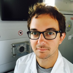
 Andrew W. Holle is a Max Planck Society Postdoctoral Fellow (2014) at the Max Planck Institute for Intelligent Systems in Stuttgart, Germany, training under Prof. Dr. Joachim Spatz of the New Materials and Biosystems department of the MPI and the Biophysical Chemistry department at Heidelberg University and Prof. Dr. Ralf Kemkemer of the Applied Chemistry department at Reutlingen University. After receiving his B.S.E. in Bioengineering at Arizona State University (2008), Dr. Holle received his Ph.D. in Bioengineering (2013) under the training of Dr. Adam Engler at the University of California, San Diego, where he studied the role of focal adhesion proteins in mechanosensitive stem cell differentiation. His current research focuses on the use of synthetic microchannels to determine the role of the cytoskeleton in ECM-dependent cancer cell migration and invasion.
