The Illinois-MGH Program in Radiological Sciences and Molecular Imaging provides faculty seminars, hosted on both the Urbana and Boston campuses. These opportunities allow for experts to share their knowledge and build relationships with colleagues located at the opposite location.
Spring 2016 Seminar Series
Dynamic and Functional Imaging of Speech and Swallowing with MRI
Brad Sutton, Ph.D., Associate Professor, Bioengineering, University of Illinois at Urbana-Champaign
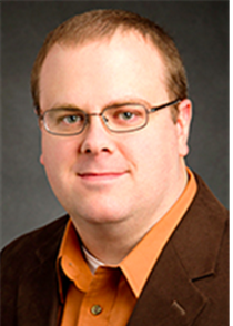 Brad Sutton, Ph.D., Bioengineering, University of Illinois at Urbana-Champaign[/caption]MRI provides several imaging contrasts to study the dynamic muscle movements and functional neural control of speech and swallowing. Leveraging a recently developed low rank imaging approach called partial separability, we have been able to achieve 150 frames per second 3D dynamic imaging of the full vocal tract during speech. This provides an unprecedented view of articulation associated with specific speech sounds. We have also developed technology to enable simultaneous monitoring of brain function and dynamic muscle movements. With these technologies, we are developing a platform for studying neuromuscular interactions in the speech and swallowing system.
Fall 2015 Seminar Series
Multimodal MRI Analysis of Speech
Jonghye Woo, Ph.D., Assistant in Physics, MGH and Assistant Professor, HMS
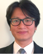 The human tongue is a volume preserving structure with highly complex, orthogonally oriented, inter-digitated muscles. This complex anatomy is necessary to move and deform the tongue throughout the vocal tract, which shapes the airway to produce swallowing and speech. However, this complex anatomy makes it extremely challenging to understand muscle specific roles and interactions among different muscles. Despite the growing need to accurately assess the similarities and differences of tongue muscular anatomy and motion patterns across different speakers, there has been a gap in our ability. Recent development in our lab of various MR technologies including high-resolution, cine, and tagged MRI allows us to investigate tongue anatomy and internal tongue motion patterns. Multimodal image analysis, based on spatio-temporal imaging data, is becoming a popular approach to relate anatomy and function in the brain and the heart. Combining imaging modalities to study tongue structure and function is an appreciable challenge from the standpoint of inferring clinically and scientifically valuable information, because its deformations are so varied.
In this presentation, he outlined our recent computational efforts toward developing state-of-the-art MR imaging and image analysis approaches to describe tongue anatomy and motion patterns during speech. Specifically, he presented methods to build a 3D vocal tract atlas and statistical models from structural MRI and a 4D atlas from cine-MRI to understand the standard anatomy and motion of the vocal tract during speech. When complete, this will be a comprehensive and systematic framework to characterize the relationship between tongue muscle structure and function during speech. Also, he presented a method to reveal functional organization of the tongue by determining functional units of the tongue motion. Taken together, the motion analysis framework has already been applied successfully to normal controls and glossectomy patients during speech.
Magnetic Nanoparticles and MR: From Imaging to Assays and Back Again
Lee Josephson, Ph.D., Associate Professor, HMS
 Lee Josephson, Ph.D., Associate Professor, MGH[/caption]The ability of Magnetic Nanoparticles (NPs) to enhance proton relaxation rates has long been recognized as a way to determine the metastatic status of normal-sized lymph nodes by MRI. Also long recognized has been the ability of magnetic NPs and MR spectrometry to serve as an in vitro assay technology. The sometimes-tortuous journey of these research concepts to commercialization, and to adaptation by the health care system, was discussed. He also discussed a recent technique for synthesizing radioactive NPs termed “taking the chemistry out of radiochemistry.” What a long strange journey the trip can be, translating nanotechnology from research to the clinic and to final societal adaptation.
Cutting-edge Radiochemical Methods and Technologies for Human PET Imaging
Neil Vasdev, Ph.D., Director of Radiochemistry, MGH and Associate Professor, HMS
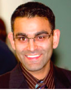 This presentation focused on some non-traditional approaches to prepare PET radiopharmaceuticals for new targets, and showed the intricacies of developing PET radiopharmaceuticals from “bench to bedside”. Specifically, cutting-edge approaches and technologies for imaging the dopaminergic pathway with PET, as well as our recent work to expand beyond the “amyloid cascade hypothesis” of Alzheimer’s disease, including tauopathies, was presented. Several of the neuroimaging agents have also been applied as oncology probes. Translating labeled compounds to PET radiopharmaceuticals and our aspiration to work towards the ultimate, albeit impossible, goal in the field: to radiolabel virtually any compound for PET was discussed.
Task Based Maximization of Information in Medical Imaging
Quanzheng Li, PhD, Physicist, MGH and Assistant Professor, HMS
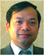 He introduced how to use signal and image processing tools to maximize the information derived from medical imaging systems for different diagnosis and prognosis tasks, and demonstrated some applications in image reconstruction (e.g. dynamic PET, time-of-flight PET and spectrum CT), image analysis (e.g. partialvolume effect correction and treatment response evaluation), and brain network analysis.
Mapping Neurotransmitter Signaling with PET and fMRI
Marc Normandin, PhD, Assistant Professor, Harvard Medical School and Assistant in Physics, MGH
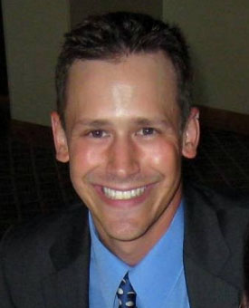 Currently available methodology for imaging neurotransmitter signaling has important limitations. While fMRI measurements of cerebral blood volume (CBV) are very sensitive to neurotransmitter release, the kinetics of the MR signal changes appear complex and often seem to lack a coherent physiological interpretation. Conventional PET methods applying targeted radioligands are often used to detect gross changes in neurotransmitter levels but these techniques ignore the transient aspect of the evoked response. In this talk, we reviewed our progress over the past decade developing PET pharmacokinetic modeling techniques that extract information about the time course of neurotransmitter levels and present a recently conceived framework for interpreting CBV-weighted fMRI in terms of the dynamics of underlying neurochemical signaling. We further described an integrated PET/fMRI kinetic modeling approach that takes advantage of simultaneous multimodal data acquisition that is possible due to the recent advent of hybrid PET/MR scanners. Applications of the techniques was discussed as well as validation studies that revealed intriguing new information about the pharmacology of drugs that were thought to be well characterized. Most of our work to date focuses on dopamine, which we used in this talk as an exemplary receptor system for more general concepts that are currently being translated to imaging the signaling of serotonin and other neurotransmitter systems.
Nanotechnology-based Approaches for Imaging and Therapy
Charalambos Kaittanis, PhD, Assistant in Chemistry MGH/HMS
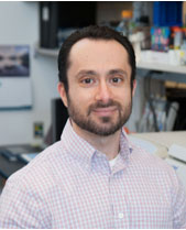 Improved diagnostics and therapeutics are needed in clinic, contributing towards better treatment and survivorship. During this presentation, we discussed how nanoparticle-based tools can support decision-making through imaging and new insights on how nanoparticles interact with their environment. Overall, we will explored how nanoparticles can serve as sensitive probes, as well as translational platforms for personalized medicine.
Spring 2015 Seminar Series
Optical Molecular Imaging of Margins and Microenvironments in Breast Cancer
Stephen A. Boppart, MD, PhD, Professor, Electrical and Computer Engineering, Bioengineering, and Medicine, University of Illinois at Urbana-Champaign
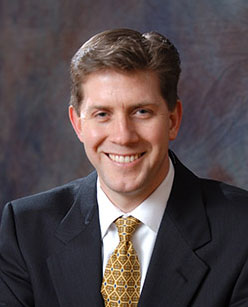 Stephen A. Boppart, MD, PhD, Professor, Electrical and Computer Engineering, Bioengineering, and Medicine, Engineering at Illinois[/caption]Advances in label-free optical imaging have enabled high-resolution imaging of breast cancer morphology and molecular composition. Real-time intraoperative optical coherence tomography has been used to assess the microstructure of breast tumor margins and lymph nodes, and new multimodal multiphoton methods can visualize the molecular tumor microenvironment. These imaging results suggest that we should also consider margins based on imaging molecular biomarkers, rather than morphological features alone.
Molecular Imaging with Spins: A Path to High Resolution through Subspaces
Zhi-Pei Liang, PhD, Professor, University of Illinois at Urbana-Champaign
 Zhi-Pei Liang, PhD, Professor, University of Illinois at Urbana-Champaign[/caption]MR spectroscopic imaging (MRSI or spatially-resolved MR spectroscopy) has been recognized as a powerful tool for label-free molecular imaging of biological systems but clinical and research applications of this technology have been developing very slowly. Conventional MRSI methods represent the desired spatio-spectral function as a vector in a very high-dimensional space. As a result, the number of spatio-spectral encodings required for decoding (or image reconstruction) can be huge, resulting in long data acquisition time, or poor spatial resolution, or low signal-to-noise ratio. It can be justified that the spatio-spectral functions of real biological systems reside in a very low-dimensional subspace. This property can be effectively utilized to accelerate MRSI experiments and achieve high resolution. This talk presented our recent advances in this exciting area of research.
Blind Compressed Sensing in Biomedical Imaging: Data-Adaptive Sparse Modeling and Acquisition
Yoram Bresler, PhD, Professor, University of Illinois at Urbana-Champaign
 Compressed sensing methods have generated tremendous interest in biomedical imaging: they can produce high quality images from significantly reduced data acquisition. We showed that replacing fixed models of sparsity and randomized acquisition in these methods by data-driven learning can help unleash the full potential of compressed sensing. These approaches are illustrated on CT and MRI.
Radiopharmaceuticals for Imaging Nuclear Receptors and Nuclear Receptor Function in Hormone-Sensitive Cancers
John A. Katzenellenbogen, Professor, Chemistry, University of Illinois at Urbana-Champaign
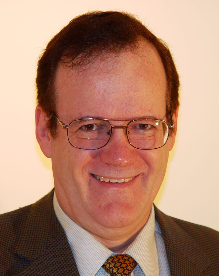 Many breast and prostate cancers are regulated by steroid receptor action, estrogen receptors and androgen receptors, respectively. We have developed fluorine-18 labeled estrogens and androgens for PET imaging the receptor contents of these cancers, and this imaging can improve the selection of patients most likely to benefit from endocrine-suppressive therapies. In addition, we are developing imaging paradigms that assess not just the presence of these receptor therapy targets, but their functional status through a hormone challenge paradigm. Functional assessment of therapy targets by longitudinal imaging can further improve and individualize the choice of optimal therapies.