Past Project: Microbial Transport in Subsurface Environment


Pathogens including Cryptosporidium parvum oocysts found in surface runoff are one of the leading causes of impaired river and estuary water. One hundred million cattle are able to produce approximately 600 oocysts annually for every person in the U.S. This number of oocysts far exceeds the infectious dose for humans. Knowledge on the fate and transport of C. parvum oocysts in agricultural runoff is currently lacking and is urgently needed to protect water supplies for many parts of the country. The results of this project will provide a scientific basis for water resources and environmental sustainability.


This project uses a multiscale approach to identify chemical and physical factors that influence attachment and mobility of C. parvum oocysts. A comprehensive understanding of these factors will be used to develop a model to predict the fate and transport of oocysts in the sub-surface environment. The objectives of this project are: (1) to investigate the role of oocyst wall macromolecules in the deposition and transport of C. parvum oocysts by systematically modifying the oocyst walls; (2) to determine the attachment mechanisms of C. parvum oocysts on inorganic (i.e. quartz) and organic (i.e. coated with natural organic matter) soil surfaces on a microscopic scale; and (3) to determine the transport of C. parvum oocysts in sub-surface environment in micromodel and column setups. The experimental approach ranges from a microscopic to a macroscopic scale. A novel microscopic technique consisting of a radial stagnation point flow (RSPF) cell combined with a microscope will be used to monitor attachment and detachment kinetics of oocysts under well-defined flow conditions in real time. Deposition and detachment experiments will be conducted with systematically varied solution conditions to determine the mechanisms of oocyst interaction with representative soil surfaces. Pore scale transport of oocyst will be studied using a precisely fabricated micromodel. Knowledge on oocyst deposition and transport at microscale will be applied to elucidate transport at continuum scale of column setup.

The main hypothesis of this project is that reversibility of oocyst deposition and subsequent transport depends on the composition of pore-water. Water contaminated with manure has significantly higher salt and organic matter than clean water such as rain. A significant portion of oocysts deposited on soil surfaces oocysts in solutions containing 3-30 mM ionic strengths is expected to detach during a sudden drop in ionic strength due to rain fall. Mobilization of the previously attached oocysts provides an additional rapid transport pathway of pathogens to water source. This hypothesis will be tested using both deposition experiments in a RSPF and transport experiments in a micromodel.
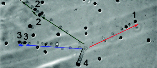
 

Microscopic observation of C. parvum oocyst deposition on the SRNOM-coated surface in secondary minima at 10 mM ionic strength. The image shown is a composite image generated by superimposing successive pictures taken over 20 min. Pictures were taken every second over the course of 30 min. Not all pictures were superimposed for clarity. C. parvum oocysts, which we traced, deposited onto the SRNOM surface and appear as single dark points marked with numbers. The paths of traced C. parvum oocysts are shown as light aligned points linked to the C. parvum oocysts marked with numbers. C. parvum oocyst 1 deposited with relatively higher velocity with an average velocity of 15.38 ?m/s (it traveled 76.9 um within 5 s); the dark point on its pathway is an oocyst that had already deposited on the SRNOM surface before C. parvum oocyst 1 appeared in the view area. C. parvum oocyst 2 was deposited 10 s after it appeared in the view area (traveling a distance of 90.1 um), released after 30 s, and deposited again 6 s later (traveling a distance of 12.5 um); eventually it was washed away by the radial flow after 95 s. C. parvum oocyst 3 was deposited 14 s after it appeared in the view area (traveling a distance of 97.4 ?m), released after 93 s, and eventually deposited 3 s later (traveling a distance of 10.2 um). C. parvum oocyst 4 deposited with a relatively lower velocity with an average velocity of 2.16 um/s (it traveled 30.2 um within 14 s). Experiments were carried out at ambient pH (around pH 5.6-5.8) and a temperature of 25 °C. Figure from Liu et al., 2009.


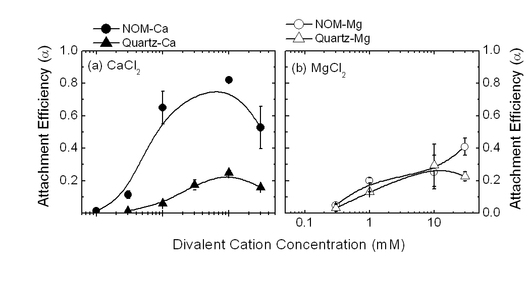
We further used a Radial Stagnation Point Flow (RSPF) system coupled with a microscope to study deposition of Cryptosporidium parvumoocysts on quartz and Suwannee River Natural Organic Matter (SRNOM)-coated surfaces in solutions with different Ca2+ or Mg2+ concentrations. Both untreated and proteinase K-treated oocysts were used. Deposition of oocysts on a SRNOM surface in Ca2+ solution was higher than in Mg2+ solution, even though the energy barriers calculated from Derjaguin-Landau-Verwey-Overbeek (DLVO) theory for Ca2+ solution were higher than for Mg2+ solution. On the other hand, the attachment of oocysts on a quartz surface was the same in both Ca2+ and Mg2+ solution and in qualitative agreement with the DLVO energy profiles. Inductive coupled plasma (ICP) was employed to measure the free divalent cation concentration in solutions containing oocysts. ICP data showed more Ca2+ bound to oocyst surface than Mg2+. Moreover, proteinase K treatment of oocysts led to a significant decrease in deposition rate due to less binding of Ca2+ to the surface of the treated oocysts as shown by the ICP data. The deposition and ICP results suggested that inner-sphere complexation of Ca2+ with carboxylate groups on both SRNOM and oocyst surfaces enhanced deposition of oocysts on a SRNOM surface.


Experimental attachment efficiencies for C. parvum oocysts on to SRNOM-coated, and quartz surfaces. Experimental conditions were as follows: capillary flow rate = 1 mL/min (average velocity) 0.0053 m/s with Re = 5.29, pH 5.6-5.8, and temperature = 25 °C. Shown data are averages and standard deviations of at least three replicates . Figure from Janjaroen et al., 2010.


We characterized the composition and conformation of Cryptosporidium parvum (C. parvum) oocyst wall surface macromolecules and studied their effect on interactions between C. parvum oocyst and quartz surface. Proteinase K and mixed glycosidases were used to modify C. parvum oocyst surface macromolecules. The peptides released by proteinase K and carbohydrates hydrolyzed by mixed glycosidases were respectively analyzed with liquid chromatography/nanoelectrospray ionization tandem mass spectrometry (LC-MS/MS) and phenol-sulfuric acid assay to determine the composition of C. parvum oocyst wall surface macromolecules. Surface potential and polarity of the untreated and proteinase treated C. parvum oocysts revealed information about the conformation of oocyst wall surface macromolecules. The results illustrated that C. parvum oocyst wall is covered by a fluffy layer of glycoproteins. Adhesion kinetics of untreated and proteinase K treated C. parvum oocysts on quartz surface were studied in a radial stagnation point flow cell over a wide range of ionic strength to investigate the effect of C. parvum oocyst wall surface macromolecules on oocysts?quartz interactions. The adhesion rate coefficient of proteinase K treated C. parvum oocysts significantly decreased compared to that of untreated oocysts. This observation indicated that the fluffy layer on C. parvum oocysts wall leads to weaker van der Waals interaction and stronger steric repulsion.

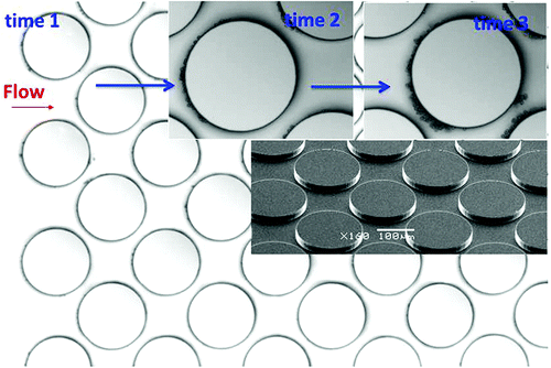
We fabricated a micromodel, which has 2-dimensional (2-D) microscopic pore structures consisting of an array of cylindrical collectors. This micromodel was used for real time monitoring of oocyst transport and attached oocyst distributions in transversal and longitudinal directions. In the micromodel, oocysts attached to the forward portion of clean collectors, where the flow velocity was lowest. After initial attachment, oocysts attached onto already attached oocysts. As a result, the collectors ripened and the region available for flow was reduced.


Real time observation of C. parvum oocysts attached to collectors in a silica micromodel. Figure from Liu et al., 2012.

Publication and Presentations

  1. Liu*, Y., Zhang, C., Hu, D., Kuhlenschmidt, M.S., Kuhlenschmidt, T.B., Mylon, S.E., Kong, R., Bhargava, R. and Nguyen, T.H., Role of Collector Alternating Charged Patches on Transport of Cryptosporidium parvum Oocysts in a Patchwise Charged Heterogeneous Micromodel. Environmental Science & Technology, 47(6), 2670-2678, 2013 full text.
  2. Liu*, Y., Zhang, C., Hilpert, M., Kuhlenschmidt, M., Kuhlenschmidt, T., Nguyen, T. H., Transport of Cryptosporidium parvum Oocyst in a silicon micromodel, Environmental Science & Technology, 2012, Vol. 46, p. 1471–1479, full text.
  3. Liu* Y., Kuhlenschmidt M. S., Kuhlenschmidt T. B., and Nguyen, T.H. , “Composition and Conformation of Cryptosporidium parvum Oocyst Wall Surface Macromolecules and Their Effect on Adhesion Kinetics of Oocysts on Quartz Surface”, Biomacromolecules, 2010, Vol. 11, pp 2109–2115, full text
  4. Janjaroen* D., Liu* Y.,Kuhlenschmidt M. S., Kuhlenschmidt T. B., and Nguyen, T.H. , “Role of Divalent Cations on Deposition Kinetics of Cryptosporidium parvum oocysts onto Natural Organic Matter Surfaces”, Environmental Science & Technology, 2010, 44, 4519–4524, full text.
  5. Liu* Y., Janjaroen* D. , Kuhlenschmidt M. S., Kuhlenschmidt T. B., and Nguyen, T.H. , “Deposition of Cryptosporidium parvum Oocysts on Natural Organic Matter Surfaces: Microscopic Evidence for Secondary Minimum Deposition in a Radial Stagnation Point Flow Cell”, Langmuir, 2009, Vol. 25, p. 1594-1605, full text
  6. Nguyen, T.H.,”Adsorption Kinetics of Cryprosporidium Parvum oocysts to Silica and Natural Organic Matter: Microscopic Evidence for Secondary Minimum Deposition”, North Carolina State University, Oct. 2008
  7. Nguyen, T.H.,”Adsorption Kinetics of Cryprosporidium Parvum oocysts to Silica and Natural Organic Matter: Microscopic Evidence for Secondary Minimum Deposition”, Swiss Federal Institute of Aquatic Science and Technology EAWAG, June 2008
  8. Liu* Y., Kuhlenschmidt M.K., Kuhlenschmidt T.K., Nguyen, T.H. Role of ionic strength on deposition kinetics of Cryptosporidium parvum oocysts to natural organic matter, presented at the 235th ACS National Meeting, New Orleans, LA, April 6-10, 2008.