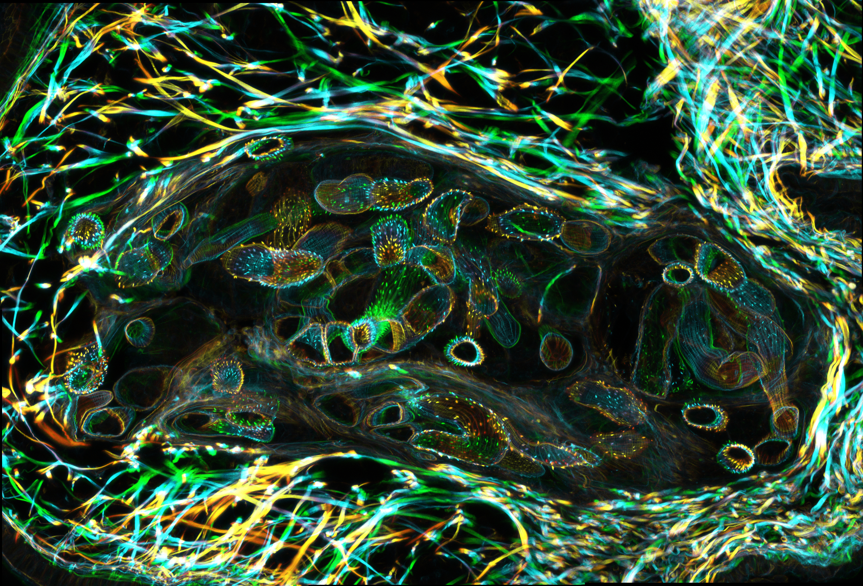
 2013. This is a cross section of a snail tentacle that harbors a parasitic flatworm called schistosome. The swirls of orange, blue, and green on the periphery of this image are snail muscle. The darker center of the image reveals a mother worm in which hundreds of daughter worms are developing. The daughters will leave the mother, migrate, and colonize other parts of the snail, and ultimately give rise to thousands even millions of individuals that can infect humans once they are released into fresh water. This massive multiplication inside the snail maximizes the transmission of this major human parasite.
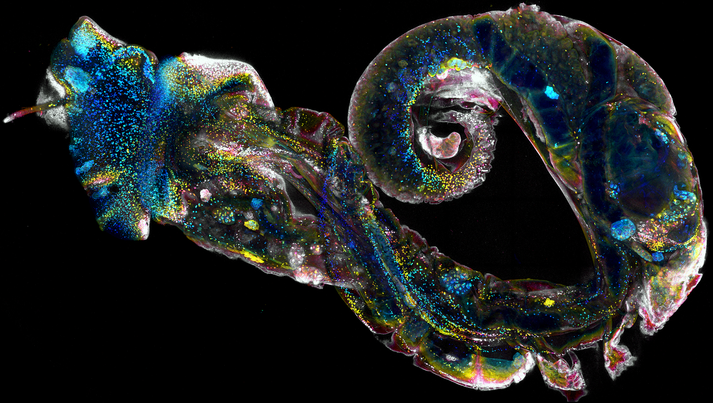 2014. This freshwater snail is infected with schistosomes. The snail was fixed, rendered optically transparent, and then imaged on a confocal microscope as a whole. Hundreds of large yellow and blue dots distributed throughout the snail body are parasites that are at various developmental stages. As these parasites mature, they will leave the snail in a few weeks to infect a human where they can live in the host vasculature for decades and lay hundreds of eggs each day. The eggs can lodge in host tissues, triggering an inflammatory response that produces extensive organ damage that can ultimately result in death. This image contains ~10 billion voxels.